
Brain
The amount of time required for full brain development is longer than any other organ. While the brain begins to develop very early in pregnancy, it grows and “fine tunes” the entire 9 months and beyond – into adulthood.
During fetal development, the brain has growth spurts and unique markers of health that can be observed via ultrasound, such as limb and facial movements. There is also evidence the fetus may begin to retain memory for sounds and language early in the third trimester.
A pregnant woman's nutrition, specifically adequate folic acid, DHA, and iodine, along with proper regulation of thyroid hormones, ensures healthy fetal brain development.
Women who have any concerns or questions regarding the development of their baby's brain (especially during/after an ultrasound), their nutrition, or their thyroid hormone status before or during pregnancy, should speak with their HCP.
Background
The length of time needed for complete brain development is significantly longer than any other organ. Brain development extends from the embryonic period into the neonatal, infant, childhood, and adolescent years. No other organ system continues to develop over such a lengthy period. Therefore, the brain develops during the entire length of pregnancy, not just the first trimester.
First Half of Pregnancy
The brain begins to develop during the 5th week of pregnancy with the formation of the 3-millimeter neural tube, which eventually develops into the brain.
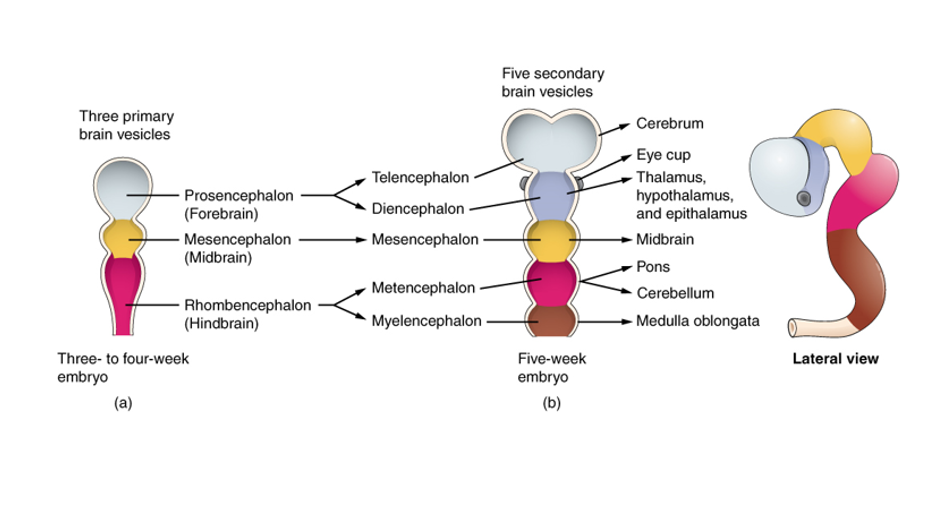
Just before neural tube closure, the end closest to where the head will eventually develop begins to expand and forms the three primary brain vesicles, or pouches. These three segments (forebrain, midbrain, hindbrain) further subdivide and by 7 to 8 weeks of pregnancy, the five secondary brain vesicles are present.
The human brainstem develops between 6 and 7 weeks of pregnancy, while the entire brain grows rapidly during week 7.
Additionally, around the same time, a mass production of brain cells develops. These cells migrate (as much as several millimeters) to very specific areas in the brain, and then become the type of cell necessary for that location. Similar cells then join up in those distinct regions, form connections, and then compete so that only the best connections remain.
The human brain begins as a smooth structure at first, and gradually develops the characteristic pattern of folding (i.e. “brain folds”). This folding of brain tissue allows large brains to fit in small skulls that also need to be small enough for the birthing process.
By 10 weeks of pregnancy, the basic structures of the brain and central nervous system are established, and the major compartments of the central and peripheral nervous systems are developed.
Central Nervous System: includes the brain and spinal cord
Peripheral Nervous System: nerves that branch off from the spinal cord and extend to all parts of the body
A newborn has about 100 billion neurons at birth; the brain must grow at the rate of about 250,000 nerve cells per minute during pregnancy. Most of these 100 billion are produced by the 20th week.
Second Half of Pregnancy
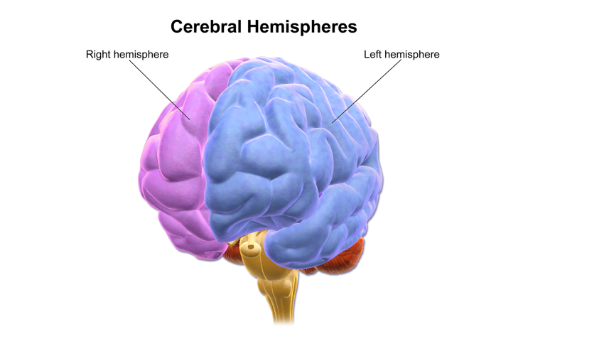
The first brain fissure to form is the fissure that separates the brain hemispheres, which develops around week 8 and is complete by week 22. (Holoprosencephaly is a birth defect that occurs when the cerebral hemispheres of the forebrain fail to divide or only partially divide. It is the most common malformation of the forebrain in humans.)
From about 17 to 22 weeks of pregnancy, the number of brain cells in the cerebral cortex increases rapidly. The cerebral cortex is the 2 to 3 mm outer covering of gray matter over the hemispheres that carries out high levels of mental functioning.
Evidence suggests that proper nutrition is of greatest importance for the development of the brain at this stage, although it also critical for the entirety of pregnancy (see Thyroid Hormones section).
By 28 weeks, fetal brainwaves can be detected through the mother's abdomen. The fetus also begins to show signs of personality and intentional behavior.
The hippocampus, which is crucial for recognition and spatial memory (learning the environment) begins growing rapidly around 32 weeks and continues after birth. The prefrontal cortex, which orchestrates complex processing behaviors, such as attention and multi-tasking, starts its growth spurt in the first six months after delivery.
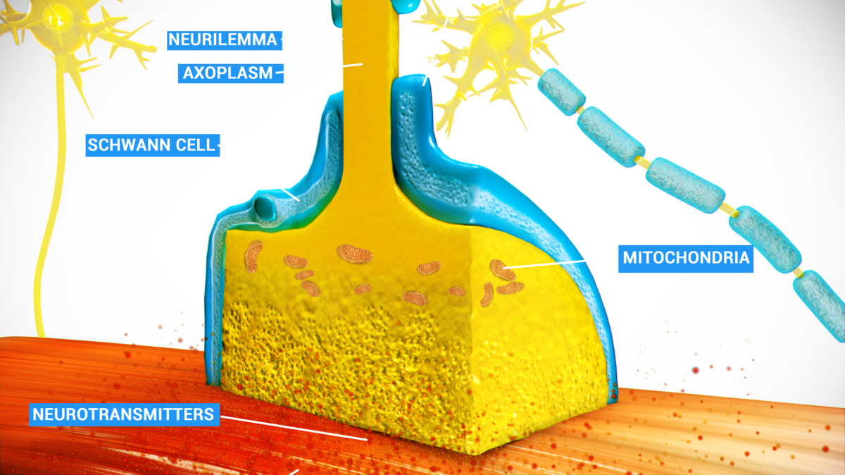
Myelination, the process in which a membrane (myelin sheath) is wrapped around axons, and enables faster flow of neural impulses throughout the brain, increases at 32 weeks. This is crucial for cognitive, motor, and sensory functions.
Myelination is assessed to be a major contributor to children’s cognitive abilities, such as processing speed, spatial working memory, and math and reading skills. Further, at least one study published preliminary findings supporting an association between myelin and processing speed in 2- to 5-year-old children.
In addition, some findings provide evidence the fetus may remember certain sounds and language beginning around 32 weeks.
Newborns appear to recognize familiar sounds and melodies, to include the sound of their mothers’ voices from other females, providing further evidence that short-term memory may be present by the end of pregnancy.
It is also possible, but somewhat unclear, if the fetus can retain memory of certain movements while in utero.
Facial movements, however, can be observed via ultrasound screenings and are indicative of healthy brain development. The fetal face, with its movements and expressions, can mirror brain function and development during different stages in utero. The face can “prove” the facial muscles and brain connections to control them, are working properly – especially when the face responds to certain stimuli outside the uterus.
The fetus’ movements also contribute to the development of the brain, as kicking, turning, and even sucking the thumb help develop and finely tune synapses.

Sound and Stress
Several studies have demonstrated that fetal exposure to excessive amounts of maternal stress can interfere with normal fetal brain development and result in harmful effects, depending on the timing, intensity, and duration of those events – with increased cortisol most likely the primary causal factor.
Cortisol, a hormone produced by the adrenal glands that has many receptors in the brain, employs a wide array of metabolic, endocrine, and immune effects. Too much cortisol could interfere with proper brain development, specifically cognitive functions.
It is also hypothesized that since positive sounds outside of the uterus help fine tune brain connections for the fetus, that an adverse prenatal sound environment may do the opposite and could have harmful effects on the brain (read Hearing).
Thyroid Hormones
Proper production of thyroid hormones by the mother is required for normal brain development of the fetus. Thyroid hormone is required early in pregnancy for the mass cell production and travel process described above. The embryo/fetus relies on the mother’s thyroid hormones the entire length of pregnancy, especially the first 18 to 20 weeks.

Maturation of the fetal thyroid gland occurs between weeks 10 and 12, with thyroid hormone production beginning around week 18.
However, the reserves of the fetal gland are low and the gland itself does not fully mature until birth, therefore the fetus still depends mostly on the mother’s thyroid hormones until birth.
Even a short period of a lack of thyroid hormones in early pregnancy creates a high risk of damage to the developing brain, which can include language and nonverbal cognitive delays. This damage could be irreversible, despite the developing brain’s ability to recover in most other cases of malnutrition.
An October 2020 study of data collected from more than 300,000 children found that when pregnant women had low levels of thyroid hormone during the first trimester, there was a 28% increase in the risk of ADHD in their offspring.
Iodine intake by the pregnant woman is critical to ensure adequate levels of thyroid hormones for fetal brain development; iodine deficiency of the mother is recognized as the most common cause of preventable fetal brain damage in the world.
Note: Pregnant women without thyroid dysfunction should be able to obtain all iodine necessary for healthy thyroid function during pregnancy by utilizing iodized salt instead of sea salt or kosher salt during pregnancy (see Iodine for more detailed information).
However, women can also have thyroid problems during pregnancy unrelated to iodine, even with no history of thyroid issues prior to pregnancy. Due to this, researchers have advocated for strict maternal thyroid hormone screening in all pregnant women by their HCPs until around week 18 to ensure normal fetal brain development. However, this screening is not routine in all practices in the United States.
Women who have any concerns or questions regarding their thyroid hormone production before or during pregnancy should speak with their HCP.
Action
Women who have any concerns or questions regarding the development of their baby's brain (especially during/after an ultrasound), nutrition, or their thyroid hormone production before or during pregnancy, should speak with their HCP.
Read Folic Acid, Neural Tube, and Iodine, and Omega-3 Fatty Acids (DHA) for more information.
Resources
New Technology Images Fetal Brain Activity in 4D (University of Washington)
Atlas of Fetal Brain MRI Images by Gestational Age (Harvard Medical School)